Diseases of crustaceans
Histological page for Yellowhead Disease
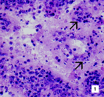 |
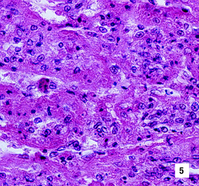
Histological section of the lymphoid organ (LO) of a juvenile giant black tiger prawn (Penaeus monodon) with severe acute yellowhead disease (YHD) at low (525x) and high (1700x) magnification. A generalised, diffuse necrosis of LO cells is shown. Affected cells display pyknotic and karyorrhectic nuclei. Single or multiple perinuclear inclusion bodies, ranging from pale to darkly basophilic, are apparent in some affected cells (arrows). This marked necrosis in acute YHD distinguishes YHD from infections due to Taura syndrome virus, which produces similar cytopathology in other target tissues, but not in the LO Source: DV Lightner |
 |
|
 |
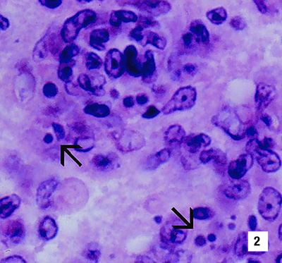
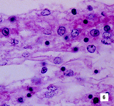
Histological section (1000x) of the gills from a juvenile black tiger prawn with YHD. A generalised, diffuse necrosis of cells in the gill lamellae is shown, and affected cells display pyknotic and karyorrhectic nuclei (arrows). A few large, conspicuous, generally spherical cells with basophilic cytoplasm are present in the section. These cells may be immature haemocytes, released prematurely in response to a YHV-induced haemocytopenia Source: DV Lightner |
 |
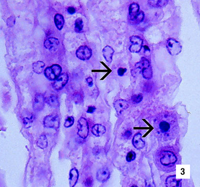
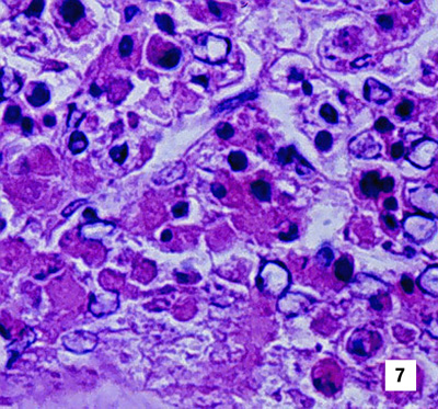
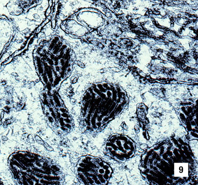
Histological sections of the LO of juvenile white shrimp (P. vannamei) (Fig 4, 1000x) and northern brown shrimp (P. aztecus) (Fig 5, 525x) experimentally infected with YHV. Severe (grade 3–4) diffuse to multifocal necrosis, characterised by cells with increased eosinophilic cytoplasm, pyknotic or karyorrhectic nuclei (arrows) and pale to densely basophilic perinuclear inclusions, is present Source: DV Lightner |
 |
|
 |
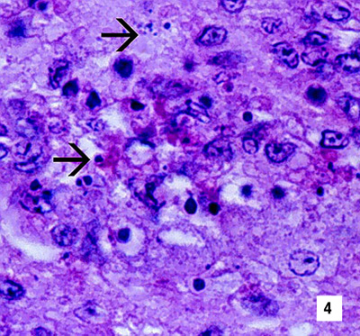
Histological sections (1000x) of the gills of a juvenile northern pink shrimp (P. duorarum) (Fig 6) and the oesophagus of a white shrimp (Fig 7) experimentally infected with YHV. Severe (grade 4) diffuse to multifocal necrosis, characterised by cells with increased eosinophilic cytoplasm, pyknotic or karyorrhectic nuclei, and pale to densely basophilic perinuclear inclusions, is present Source: DV Lightner |
 |
|
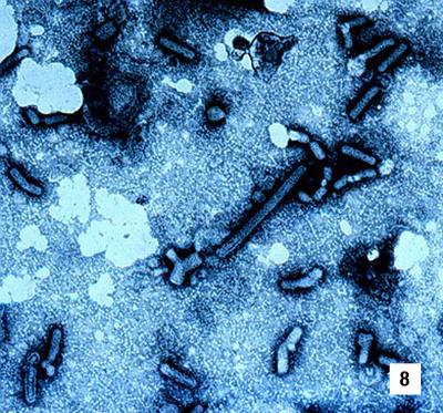 |
Semipurified preparation of YHV from the haemolymph of an experimentally infected white shrimp. Rod-shaped, enveloped virions of YHV, which are highly variable in length, are illustrated. 2% PTA stain, ~18 000x Source: DV Lightner |
 |
TEM (~50,000x) of a section of YHV-infected tissue from a giant black tiger prawn. Cytoplasmic inclusions packed with enveloped virions of YHV are well shown. Less obvious but present near the membrane-bound viral inclusions are very thin, fibre-like to rod-shaped, unenveloped nucleocapsids of YHV. (Micrograph: TW Flegel, Bangkok, Thailand) Source: DV Lightner |