Diseases of crustaceans
Histological page for White spot Disease
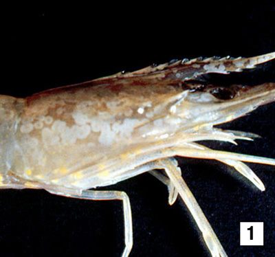 |
A juvenile giant black tiger prawn (Penaeus monodon) that is displaying the distinctive white spots of white spot disease (WSD). White spots are especially visible on the carapace and the rostrum. While providing a tentative diagnosis of WSD infection, white spots are not always visible in shrimp with acute phase white spot syndrome, and may develop in the subacute to chronic or recovery phases of the infection Source: DV Lightner |
|---|---|
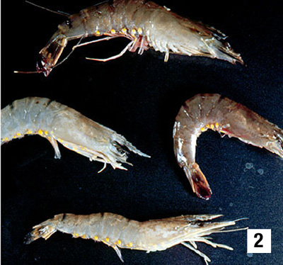 |
Four juvenile black tiger prawns, including the one shown in Fig 1 (at bottom), with different gross signs of white spot syndrome. The top and right shrimp show few, if any, white spots, but show a pink to red-brown discolouration due to expansion of the subcuticular chromatophores. This reddish appearance may be a gross sign that is more apparent in the acute phase of the disease. The shrimp on the left and bottom display diagnostic white spots that develop after the acute phase of the disease Source: DV Lightner |
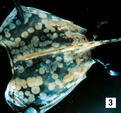 |
The carapace from a juvenile black tiger prawn with WSD. Calcareous deposits on the underside of the shell account for the white spots. Photo: P Saibaba, SKBR College, Amalapuram, India Source: DV Lightner |
 |
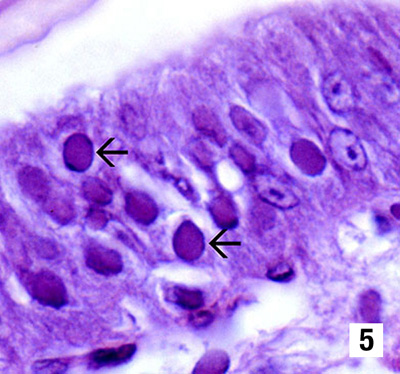
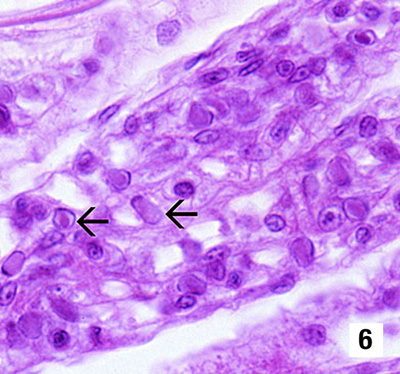
Photomicrograph (900x) of a histological section from the stomach of a juvenile black tiger prawn infected with WSD. Prominent intranuclear inclusion bodies are abundant in the cuticular epithelium and subcuticular connective tissue of the organ (arrows). Cells in different phases of infection by WSD display intranuclear inclusion bodies. The early phase inclusion bodies that predominate in this section are centronuclear, eosinophilic, and separated from the nuclear membrane and marginated chromatin by an artifactual halo (resembling infectious hypodermal and haematopoietic necrosis inclusion bodies) Source: DV Lightner |
 |
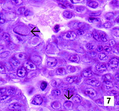
Histological section (1300x) of the stomach of a juvenile Chinese white shrimp (P. chinensis) with an advanced WSD infection. Fully developed WSD intranuclear inclusion bodies (arrows) are more basophilic, appear granular in texture, and nearly fill the affected hypertrophied nucleus. Occlusion bodies are not present Source: DV Lightner |
 |
Section of the gills from a juvenile Chinese white shrimp with WSD (900x). Nearly one-quarter of the cells present are infected, as indicated by the presence of developing and fully developed intranuclear inclusion bodies of WSD (arrows) Source: DV Lightner |
 |
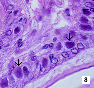
Section of a WSD-infected haematopoietic nodule (1300x) from a juvenile Chinese white shrimp. As in Fig 6, nearly one-fourth of the cells present in the section display intranuclear inclusions bodies of WSD (arrows) in various stages of development Source: DV Lightner |
 |
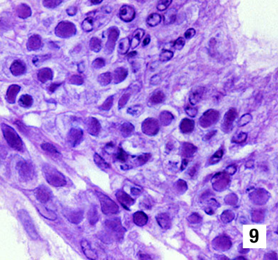
Histological sections (900x) of the stomachs of blue shrimp (P. stylirostris, Fig 8) and white shrimp (P. vannamei, Fig 9) experimentally infected with WSD. Both species display severe (grade 4) infections by WSD, with classic WSD intranuclear inclusion bodies (arrows) that are identical to those illustrated in Figs 3–7 Source: DV Lightner |
 |
|
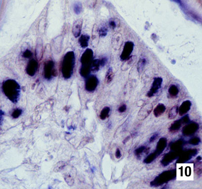 |
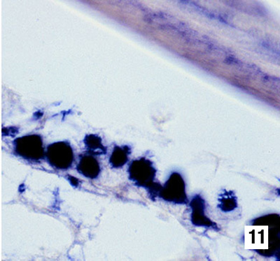
Sections of various tissues from a WSD-infected juvenile white shrimp reacted by in situ hybridisation with a DIG-labelled DNA probe to the virus. The probe has reacted strongly with intranuclear inclusion bodies containing WSD in the various tissues of this shrimp, including the cuticular epithelium of the stomach (Fig 10, 900x), the cuticular epithelium and connective tissues of the carapace (Fig 11, 900x), and epithelial cells in the antennal gland (Fig 12, 450x) Source: DV Lightner |
 |
|
 |