Diseases of crustaceans
Histological page for Taura Syndrome
 |
Moribund, juvenile, pond-reared white shrimp (Penaeus vannamei) from Ecuador in the peracute phase of Taura syndrome (TS). The shrimp are lethargic, have soft shells, and a distinct red tail fan Source: DV Lightner |
|---|---|
 |
A higher magnification (10x) view of the tail fan of one of the two shrimp shown in Fig 1. Use of a hand lens (or the close-up lens on a camera) shows rough edges of the cuticular epithelium in the uropods that are suggestive of focal necrosis of the epithelium at those sites (arrow) Source: DV Lightner |
 |
Juvenile, pond-reared white shrimp (Fig 3 from Ecuador and Fig 4 from Texas) in the chronic or recovery phase of TS. Multiple melanised foci mark sites of resolving cuticular epithelium necrosis due to TSV infection Source: DV Lightner |
 |
|
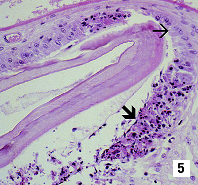 |
A histological section through the stomach of a juvenile white shrimp with peracute TS. Prominent areas of necrosis in the cuticular epithelium (large arrow), which secretes the overlying acellular cuticle, are apparent. Adjacent to the focal lesions are normal looking epithelial cells (small arrow). 300x Source: DV Lightner |
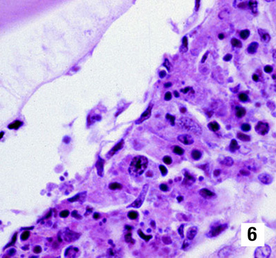 |
A higher magnification (900x) of one of the classic peracute phase TS lesions shown near the centre of Fig 5. Classic TS lesions consist of necrotic cuticular epithelial and subcuticular connective tissue cells with pyknotic and karyorrhectic nuclei, a generally increased cytoplasmic eosinophilia, and very numerous, variably staining, cytoplasmic inclusions. The cytoplasmic inclusions and pyknotic and karyorrhectic nuclei give the lesion a pathodiagnostic 'peppered' or 'buckshot-riddled' appearance. The peracute nature of the lesion is suggested by the absence of haemocytes in or near the lesion Source: DV Lightner |
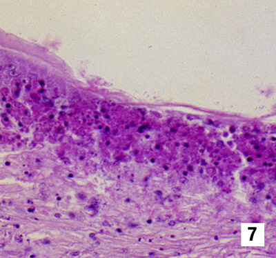 |
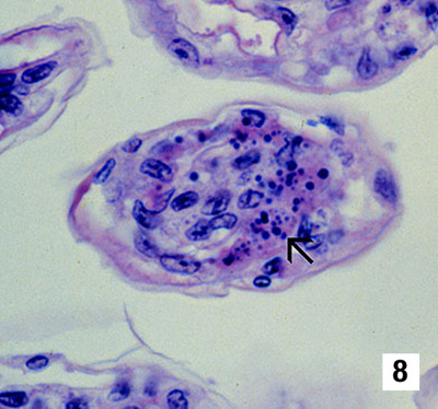
Pathognomonic focal TSV lesions in tissues (other than those shown in Figs 5 and 6) of a juvenile white shrimp with peracute TS. Fig 7 (450x) is a lesion in the cuticular epithelium and subcutis of the carapace; Fig 8 (900x) is in the gills (arrow). Nuclear pyknosis and karyorrhexis, increased cytoplasmic eosinophilia, and an abundance of variably staining, generally spherical cytoplasmic inclusions are distinguishing characteristics of the lesions Source: DV Lightner |
 |
|
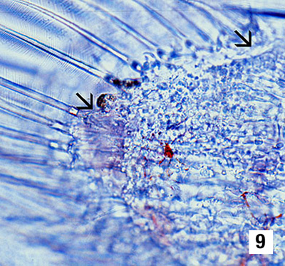 |
Wet-mount of a uropod of an experimentally infected postlarval white shrimp in the peracute phase of TS. The postlarva was in the D4 stage of its moult cycle, as shown by the presence of the 'old' cuticle separated from the 'new' cuticle by a space. The arrows mark the approximate margins of a focal area of necrosis in the cuticular epithelium. The area of necrosis is evidenced by the presence of a vacant zone just under the cuticular epithelium (where the cuticular epithelium should be) and by the presence of refractile spheres (which are pyknotic and karyorrhectic nuclei) near the periphery of the lesion. A few expanded red chromatophores are also apparent in the subcuticular connective tissues of the uropod. No stain, 300x Source: DV Lightner |
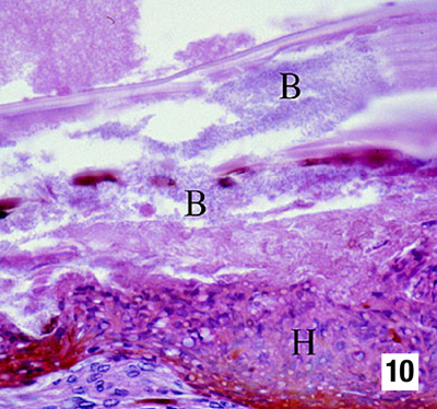 |
Histological section (600x) of a resolving cuticular lesion in a juvenile white shrimp. A perforated cuticle that is heavily colonised with masses of bacteria (B) is at the top of the micrograph. A thick, melanised, haemocytic 'plug' (H) has formed basal to the cuticular epithelium to temporarily close the 'wound' from the outside. Basal to the haemocyte plug (H), connective tissue elements, and additional infiltrating haemocytes, provide the basal support for the regeneration of the cuticular epithelium. Pathognomonic TS lesions in the recovery/chronic phase of TS are usually few, relative to the resolving lesions shown here, and are often entirely absent. Source: DV Lightner |
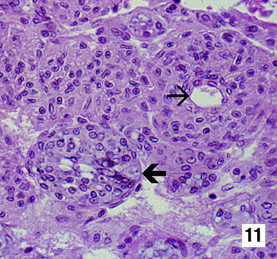 |
Mid-sagittal section (450x) of the lymphoid organ (LO) of an experimentally infected juvenile white shrimp in the chronic or recovery phase of TS. While pathognomonic TS lesions of the type seen in the cuticular epithelium never occur in the LO, TSV does induce some significant lesions in this organ. Interspersed among normal looking LO cords or tissue, which is characterised by multiple layers of sheath cells around a central hemolymph vessel (small arrow), are accumulations of disorganised LO cells that form LO 'spheroids' (LOS). LOS lack a central vessel and consist of cells that show karyomegaly and large prominent cytoplasmic vacuoles and other cytoplasmic inclusions (large arrow) Source: DV Lightner |
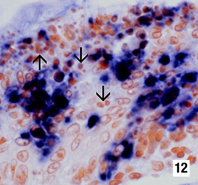 |
Histological section (900x) of an appendage from a postlarval white shrimp in the peracute phase of TS that has been reacted with a DIG-labelled cDNA probe to TSV. The probe has reacted intensely with TSV-infected cells, staining the cytoplasm of infected cuticular epithelial cells and subcuticular connective tissue cells positive for the virus. The probe does not react with the pyknotic and karyorrhectic nuclei (arrows), which is not unexpected because TSV is only cytoplasmic. These nuclear remnants contribute to the 'peppered' or 'buckshot-riddled' appearance of TS lesions Source: DV Lightner |