Diseases of crustaceans
Histological page for Tetrahedral Baculovirosis
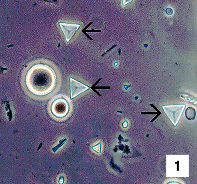 |
Wet-mount of faeces from a white shrimp (Penaeus vannamei) with tetrahedral baculovirosis. The tetrahedral occlusion bodies (TOBs; arrows) are diagnostic for infection of the shrimp's hepatopancreas (HP) or midgut epithelial (MG) cells. TOBs are released into the gut contents by the necrosis and lysis of tetrahedral baculovirus-infected HP or MG epithelial cells. 700x Source: DV Lightner |
|---|---|
 |
Low (Fig 2, 350x) and mid (Fig 3, 700x) magnification views of mid-sagittal sections of postlarva white shrimp with severe (grade 3–4) tetrahedral baculovirus infections of the HP. Baculovirus-infected cells display multiple eosinophilic baculovirus TOBs within markedly hypertrophied HP cell nuclei (arrows) Source: DV Lightner |
 |
|
 |
High-magnification (1800x) photomicrograph of an HP tubule showing several tetrahedral baculovirus-infected cells that illustrate well the diagnostic intranuclear, eosinophilic, tetrahedral (triangular or rhombohedral in section) occlusion bodies of baculovirus (arrows) Source: DV Lightner |
 |
Low-magnification (150x) view of a tetrahedral baculovirus-infected postlarva white shrimp that is similar in age and infection severity to the postlarva shown in Fig 2, but reacted with a DIG-labelled DNA probe for tetrahedral baculovirus. Baculovirus-infected cells are stained dark blue by the probe. Note that infected cells are confined to the HP and MG, and that baculovirus-positive cells are not present in the surrounding non-enteric tissues. Some nonspecific staining of the cuticle by the probe is apparent Source: DV Lightner |
 |
High-magnification (700x) photomicrograph of the HP of a juvenile white shrimp infected with tetrahedral baculovirus. The section was reacted with a DIG-labelled DNA probe. Intact infected HP cell nuclei provide an intense positive reaction for virus and viral DNA that is free within the nucleoplasm (large arrow). However, because the TOBs are not penetrated by the probe, the TOBs by themselves do not show a positive reaction for the virus despite their viral content (small arrow) Source: DV Lightner |