Diseases of crustaceans
Histological page for Spherical Baculovirosis
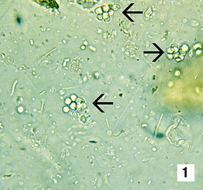 |
A wet-mount prepared from the midgut contents of an adult giant black tiger prawn (Penaeus monodon) infected with spherical baculovirosis. Clusters of spherical occlusion bodies (arrows), held together by remnants of the nuclear membrane, are illustrated. No stain, 700x Source: DV Lightner |
|---|---|
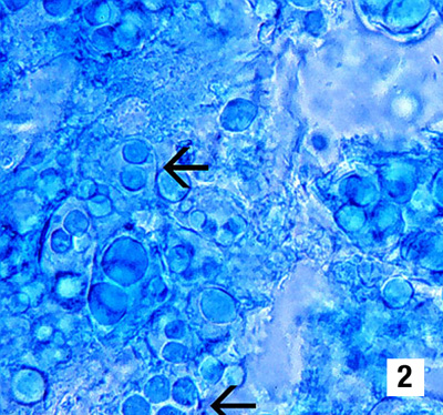 |
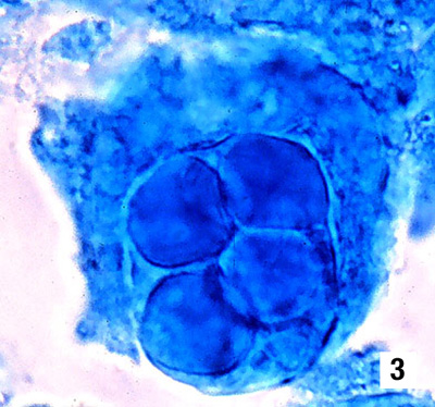
Mid (Fig 2, 700x) and high (Fig 3, 1700x) magnification views of tissue squash preparations of the hepatopancreas (HP) from postlarval (PL) black tiger prawns with grade 4 spherical baculovirus infections. Most HP cells in both PLs display multiple, generally spherical, intranuclear occlusion bodies (arrows) that are diagnostic for spherical baculovirosis. 0.1% malachite green Source: DV Lightner |
 |
|
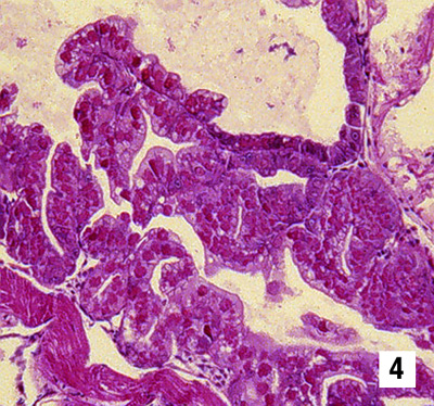 |
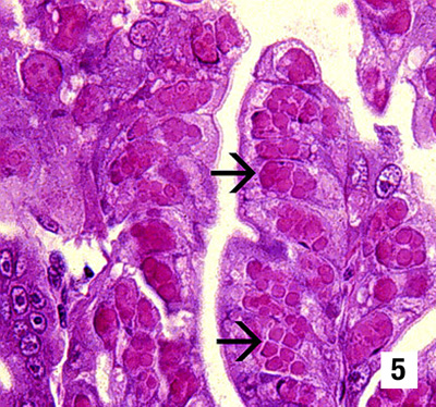
Low (Fig 4, 175x) and high (Fig 5, 700x) magnification views of the same mid-sagittal section of the hepatopancreas of a PL black tiger prawn with a grade 4 spherical baculovirus infection. Only one HP tubule in the lower left of Fig. 5 contains normal looking HP cells. Virtually all of the remaining HP tubules are displaying infected cells with fully developed intranuclear occlusion bodies. In H&E preparations, spherical baculovirus occlusion bodies appear as eosinophilic, generally multiple, spherical inclusion bodies in enormously hypertrophied nuclei (arrows) Source: DV Lightner |
 |
|
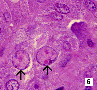 |
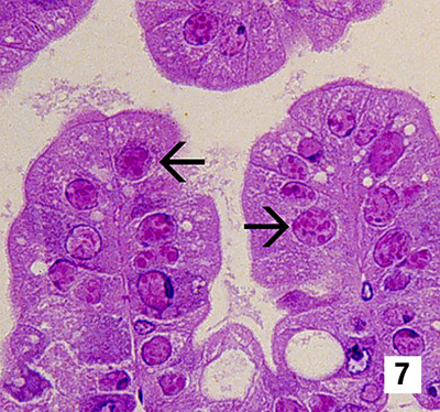
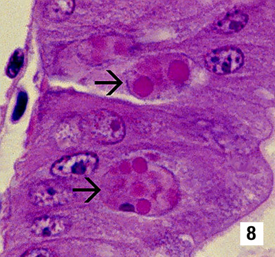
Mid (700x) and high (1700x) magnification views of spherical baculovirus-infected cells in the HP of PL or juvenile black tiger prawn. Different stages of development are illustrated in the photomicrographs, ranging from very early to fully developed. Fig 6 best illustrates an early stage of infection when nuclear hypertrophy and chromatin margination have occurred, but occlusion body formation has just begun. Figs 7 and 8 show all stages of spherical baculovirus infection development Source: DV Lightner |
 |
|
 |
|
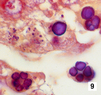 |
Spherical baculovirus occlusion bodies may be stained with a variety of stains, such as tissue Gram stains for bacteria. In this example, baculovirus occlusions have been stained purple. However, the results are highly variable and red staining with tissue Gram stains also occurs. Brown and Brenn Gram stain, 1700x Source: DV Lightner |
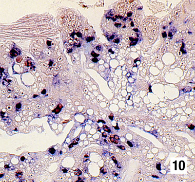 |
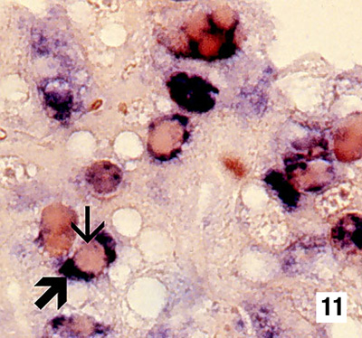
Low (Fig 10, 250x) and high (Fig 11, 1000x) magnification views of a mid-sagittal section of the HP from a spherical baculovirus-infected black tiger prawn, which has been reacted with a DIG-labelled DNA probe to the virus. Spherical baculovirus occlusion bodies are impermeable to the baculovirus probe (small arrow), but they are surrounded by intensely baculovirus-positive intranuclear areas, as marked by the dark blue stain (large arrow). DIG-labelled probe and Bismarck Brown Source: DV Lightner |
 |