Diseases of crustaceans
Histological page for Necrotising Hepatopancreatitis
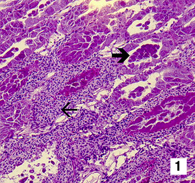 |
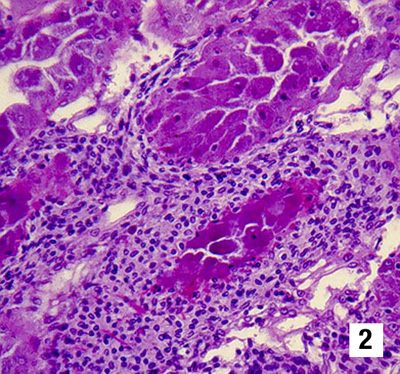
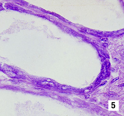
Low (Fig 1, 150x) and mid (Fig 2, 300x) magnification photomicrographs of the hepatopancreas (HP) of a juvenile white shrimp (Penaeus vannamei) with severe, subacute (grade 3–4) necrotising hepatopancreatitis (NHP). Severe haemocytic inflammation (with some melanised foci) of the intratubular spaces (small arrow) in response to necrosis, cytolysis, and sloughing of HP tubule epithelial cells (large arrow) are among the principal histopathological changes due to NHP Source: DV Lightner |
|---|---|
 |
|
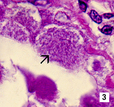 |
A high magnification (1700x) view of a portion of the HP from Figs 1 and 2. The HP tubule epithelial cells show no cytoplasmic lipid droplets, but instead contain masses of the tiny, non-membrane bound, intracytoplasmic NHP bacteria (arrow) Source: DV Lightner |
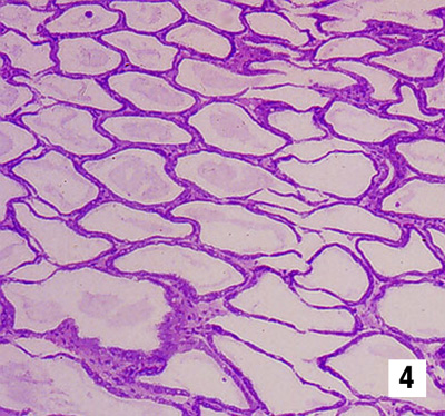 |
Low magnification (100x) view of the HP of a juvenile white shrimp with severe, chronic NHP. The HP tubule epithelium is markedly atrophied, resulting in the formation of large oedematous (fluid filled or 'watery') areas in the HP Source: DV Lightner |
 |
A higher magnification (900x) photomicrograph of the atrophied HP from a juvenile white shrimp with chronic NHP. In contrast to the subacute phase of NHP, chronic phase HPs shown no, or only occasional, foci of haemocytic inflammation (large arrow) of the necrotic or degenerating HP tubules. NHP bacteria (small arrow) may be found in the cytoplasm of an occasional HP cell Source: DV Lightner |
 |
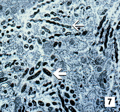
Section of the HP of a juvenile white shrimp that is similar to that shown in Fig 3. Cytoplasmic masses of the NHP bacterium are silver stained and appear brown to black with the modified Steiner stain. Unaffected cells and nuclei are pale brown (1600x) Source: DV Lightner |
 |
TEM (10 000x) of a hepatopancreatocyte from a juvenile white shrimp with NHP. Profiles of intracellular rod-shaped forms (large arrow) and helical forms (small arrow) of the NHP bacterium are abundant in the cytoplasm Source: DV Lightner |
 |
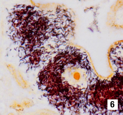
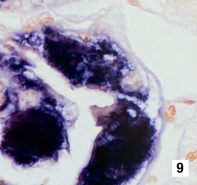
Low (Fig 8, 250x) and high (Fig 9, 1000x) magnification views of sections of the HP of a juvenile white shrimp with NHP. This section has been assayed for NHP using a DIG-labelled DNA probe. Cytoplasmic masses of the NHP bacterium are marked blue to blue-black by the probe. Unaffected cells and host cell nuclei take the brown counter stain. DIG-labelled NHP probe and Bismarck Brown Source: DV Lightner |
 |