Diseases of finfish
Histological page for Bacterial kidney disease
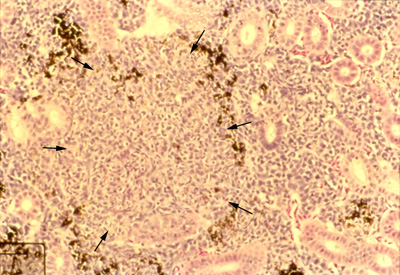 |
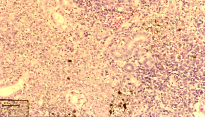
Focal (top photo, arrows) and diffuse (bottom photo) granulomas in the posterior kidney of juvenile chinook salmon Oncorhynchus tshawytscha with BKD. Hematoxylin and eosin stain; scale bars = 50 µm. Photos courtesy of Dr. Caroline O’Farrell. Source: R Pascho/C O'Farrell |
|---|---|
 |
|
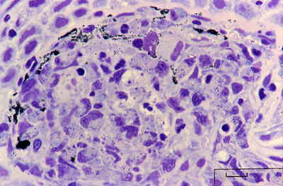 |
Histological section of a skin lesion of a juvenile chinook salmon Oncorhynchus tshawytscha infected with Renibacterium salmoninarum. Most of the small rod-shaped R. salmoninarum are visible within the cytoplasm of macrophages. Note that the bacteria are purple-blue in this Giemsa-stained preparation, in contrast to the black melanin granules. Source: R Pascho |
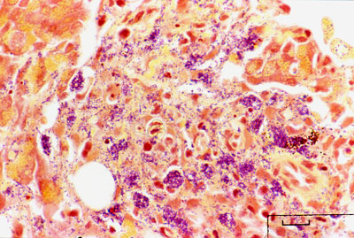 |
Gram-stained histological section of pancreatic tissue of a juvenile chinook salmon Oncorhynchus tshawytscha with systemic BKD. Renibacterium salmoninarum cells are present extracellularly and intracellularly within macrophages. Note the color difference between the gram-positive (purple-blue) bacteria and the brown-black melanin granules (arrow, inset). Source: R Pascho |