Diseases of molluscs
Histological page for infection with with Mikrocytos mackini
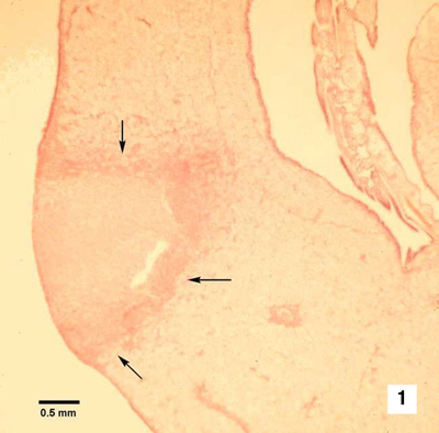 |
Histological section through a lesion caused by Mikrocytos mackini on the mantle of Pacific oyster (Crassostrea gigas). This intracellular protozoan (not visible at this magnification) usually occurs in the intact vesicular connective tissue cells immediately surrounding the periphery of the lesion (arrows). Haematoxylin and eosin stain
Source: S Bower |
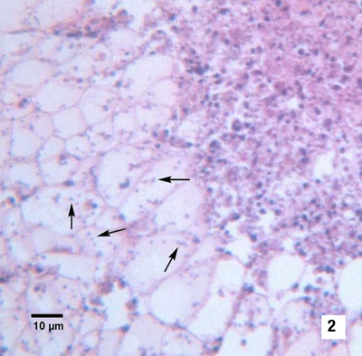 |
Many M. mackini (arrows) within vesicular connective tissue cells adjacent to a lesion characterised by an accumulation of haemocytes and necrotic cells. Haematoxylin and eosin stain Source: S Bower |
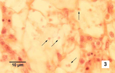 |
Oil immersion magnification (100 x objective) of M. mackini (arrows) within the cytoplasm of vesicular connective tissue cells of Pacific oyster. Haematoxylin and eosin stain Source: S Bower |
 |
As for Fig 3 but from a different specimen. Because of the small size of this parasite, it is very difficult to visualise and photograph in histological preparations. Haematoxylin and eosin stain Source: S Bower |
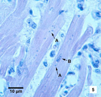 |
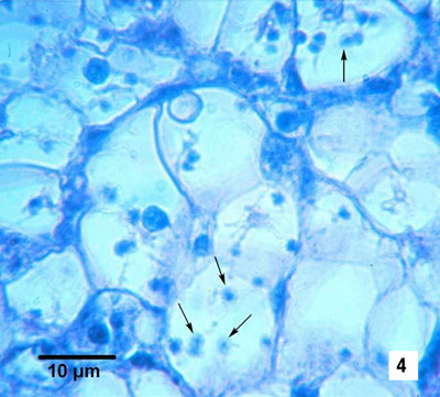
M. mackini (A) within fibres of the adductor muscle of Pacific oyster. One M. mackini is located close to the nucleus (B) of a muscle cell. Haematoxylin and eosin stain Source: S Bower |
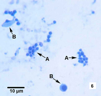 |
Clusters of M. mackini (A), isolated and partially purified from a heavily infected Pacific oyster, among host cell debris (B). Hemacolor® stain Source: S Bower |
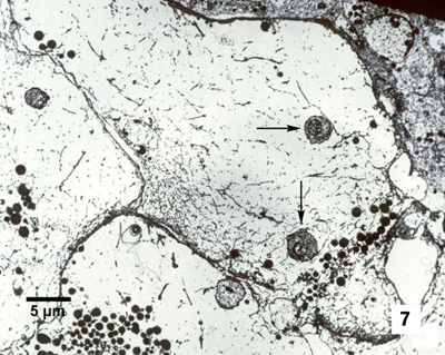 |
Electron micrograph of a Pacific oyster vesicular connective tissue cell containing M. mackini (arrows). Uranyl acetate and lead citrate stain Source: S Bower |
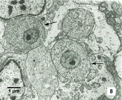 |
M. mackini (arrows) each containing a nucleus with a pronounced nucleolus and lacking mitochondria. Uranyl acetate and lead citrate stain Source: S Bower |
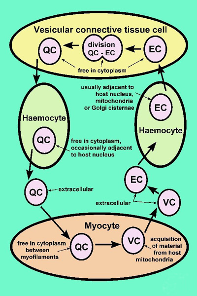 |
Proposed developmental cycle of
M. mackini indicating host cell type and host organelle affiliation
for the three recognised morphological forms: quiescent cell (QC), vesicular
cell (VC) and edosomal cell (EC) Source: S Bower |