Diseases of molluscs
Parasitic diseases—Infection with Mikrocytos mackini
CLICK ON IMAGE TO ENLARGE
Source: S Bower
CLICK ON IMAGE TO ENLARGE
Source: S Bower
CLICK ON IMAGE TO ENLARGE
Source: S Bower
Signs of disease
Important: animals with disease may show one or more of the signs below, but disease may still be present in the absence of any signs. Macroscopic lesions are not always present.
Gross signs of disease in an infected animal
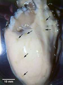
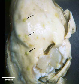
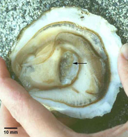
- focal green pustules up to 5 mm in diameter within body wall or on surfaces of the labial palps or mantle
- brown scars often on shell adjacent to abscesses or ulcers on the mantle surface
- focal intracellular infection, mainly of vesicular connective tissue cells, resulting in haemocyte infiltration and tissue necrosis
- abscesses composed of granular haemocytes and hyalinocytes, and may contain small (1–3 µm) cells
Disease agent
Mikrocytos mackini, a member of the phylum Haplosporidia, is a parasite of connective tissue that causes lethal infection of the haemocytes of certain oysters.
Host range
Molluscs known to be susceptible to infection with M. mackini:
European flat oyster* (Ostrea edulis)
Olympia oyster* (Ostrea conchaphila)
Pacific oyster* (Crassostrea gigas)
American oyster (Crassostrea virginica)
* naturally susceptible (other species have been shown to be experimentally susceptible)
Presence in Asia–Pacific
EXOTIC — has not been officially reported in the Asia–Pacific region under the NACA–FAO–OIE quarterly aquatic animal disease reporting program.
Epidemiology
- Severe infections appear to be restricted to oysters over two years old.
- The mortality rate varies at around 40% of older oysters at low tide levels.
- The disease occurs more often in April and May (the Northern Hemisphere spring), after a 3–4 month pre-patent period when temperatures are less than 10°C.
- Mikrocytos mackini prefers high salinities.
- The Pacific oyster seems to be more resistant to the disease than the other species challenged experimentally, under laboratory and field conditions.
Differential diagnosis
The differential diagnostic table and the list of similar diseases appearing at the bottom of each disease page refer only to the diseases covered by this field guide. Gross signs observed might well be representative of a wider range of diseases not included here. Therefore, these diagnostic aids should not be read as a guide to a definitive diagnosis, but rather as a tool to help identify the listed diseases that most closely account for the gross signs.
Outside the known distribution range, electron microscopy or molecular probes (if available) must be used to identify and distinguish the detected organism from microcell species of Bonamia.
Sample collection
Because of uncertainty in differentiating diseases using only gross signs, and because some aquatic animal disease agents might pose a risk to humans, you should not try to collect samples unless you have been trained. Instead, you should phone your national hotline number and report your observations. If samples have to be collected, the agency taking the call will advise you on what you need to do. Local or district fisheries/veterinary authorities could advise you on sampling.
Emergency disease hotline
For your national emergency disease hotline number, see Whom to contact if you suspect a disease.
Further reading
http://www.oie.int/aac/eng/cards/en_diseasecard.htm
http://www.pac.dfo-mpo.gc.ca/sci/shelldis/pages/mikmacoy_e.htm
The currently accepted procedures for a conclusive diagnosis of infection with Mikrocytos mackini are summarised at http://www.oie.int/eng/normes/fmanual/A_00041.htm
These hyperlinks were correct and functioning at the time of publication.