Diseases of molluscs
Histological page for infection with Marteilia sydneyi (QX Disease)
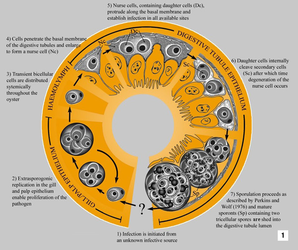 |
Hypothetical development of Marteilia sydneyi in Sydney rock oyster (Saccostrea glomerata) Source: S Kleeman |
|---|
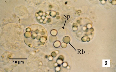 |
Sporonts of M. sydneyi containing refractile bodies (Rb) and spores (Sp) Source: S Kleeman |
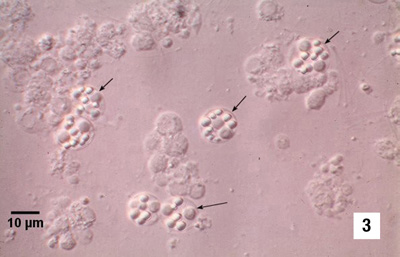 |
Sporonts of M. sydneyi (arrows) viewed under interference contrast optics Source: S Kleeman |
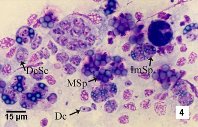 |
Hemacolor (Merck) stained tissue imprint of the digestive gland of Saccostrea glomerata infected with M. sydneyi, showing various lifecycle stages including daughter cells (Dc), daughter cells containing secondary cells (DcSc), immature sporonts (ImSp), and mature sporonts (MSp). Note that the various stages observed are often ruptured from their enclosing cells (ie the nurse cells or sporangiosori) Source: S Kleeman |
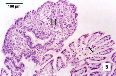 |
Oyster reaction consisting of epithelial and connective tissue hyperplasia (H) and fusion of filaments due to the presence of numerous extrasporogonic stages in the epithelium of the gills, in contrast to relatively normal-looking gill tissue (N) Source: S Kleeman |
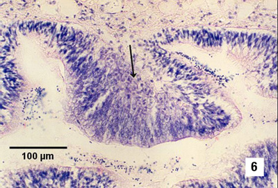 |
Replicating stages in the palp epithelium. Note the hypertrophy of the epithelial cells in the presence of proliferating parasites (arrow) in infected areas Source: S Kleeman |
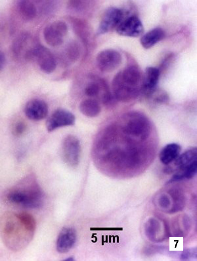 |
Higher magnification of extrasporogonic stages in the epithelium of the gills (see phase 2 in Fig 1) Source: S Kleeman |
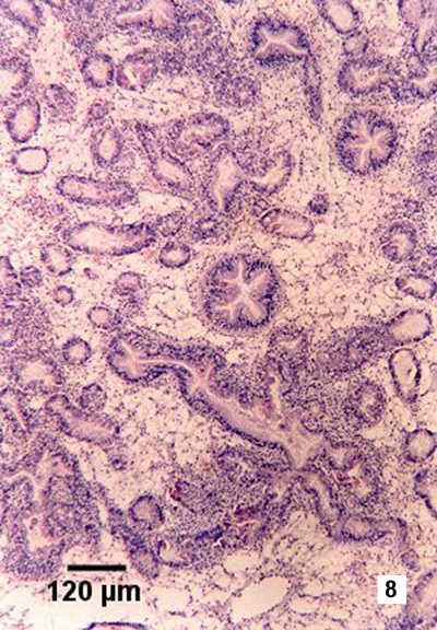 |
Section showing haemocytic infiltration of the connective tissue surrounding infected digestive gland tubules. Haematoxylin and eosin stain Source: S Kleeman |
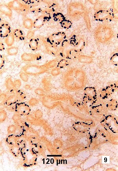 |
Tissue section showing the location of presporulating nurse cell stages (stained black) in digestive gland tubule epithelia Source: S Kleeman |
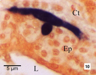 |
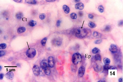
Nurse cell (stained black by in situ hybridisation) demonstrating the extent of the pseudopodial extensions along the basal membrane of the digestive tubule epithelium (Ep). This feature is not evident with haematoxylin and eosin staining. Other labelled features are the connective tissue (Ct) that surrounds the tubule and the lumen (L) of the tubule Source: S Kleeman |
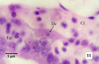 |
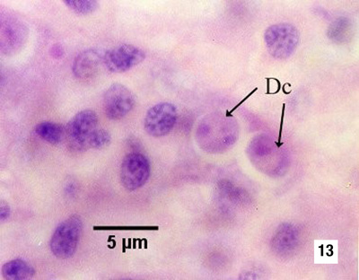
Nurse cell containing one daughter cell (Dc) and residing along the basal membrane of the tubule between the connective tissue (Ct) surrounding the tubules and the tubule epithelium (Ep) Source: S Kleeman |
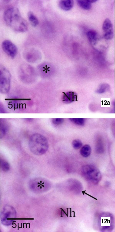 |
The same tissue section but at different focal planes demonstrates the budding of a daughter cell (arrow in Fig 12b) within the nurse cell. An asterisk marks the same daughter cell and Nh denotes the same host cell nucleus in each figure. There are two additional daughter cells within the nurse cell (Fig 12a) Source: S Kleeman |
 |
Nurse cell containing two daughter cells (Dc, see phase 5 in Fig 1) Source: S Kleeman |
 |
Nurse cells containing bicellular daughter cells (arrows) along the basal membrane between the tubule epithelium (Ep) and the connective tissue that contains many infiltrating haemocytes (see phase 6 in Fig 1) Source: S Kleeman |
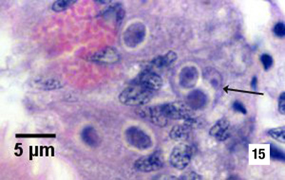 |
Primary cell (arrow) containing two secondary cells (sporont primordia) just before sporulation (see initiation of phase 7 in Fig 1) Source: S Kleeman |
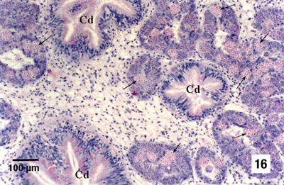 |
Numerous sporulating stages (arrows) in the digestive gland tubules. Note that sporulation does not occur in the ciliated ducts (Cd) of the digestive gland Source: S Kleeman |
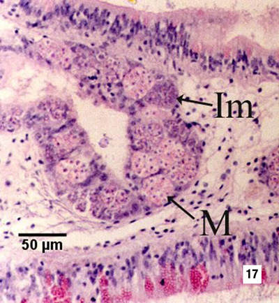 |
Immature sporonts (Im) and mature sporonts (M) within sporangiosori in a digestive gland tubule. Note that the epithelium of the tubule is almost completely replaced by M. sydneyi Source: S Kleeman |